Laboratorio de Investigación en Cultivo Celular e Inmunología Molecular

El laboratorio investiga compuestos bioactivos de plantas medicinales y macroalgas usando cultivos de células cancerosas. Además, aísla células madre de diversas especies animales y líneas celulares cancerosas para estudiar exosomas, y evalúa la respuesta inmunitaria contra el SARS-CoV-2 en pruebas ex vivo.

Jefe del laboratorio

Ana Lucía Mayanga Herrera

Personal de apoyo

Daniella Fernanda Orihuela Castillo -Investigadora Asistente
Marcos Antonio Echenique Carhuajulca – Auxiliar Técnico de Laboratorio

Líneas de investigación

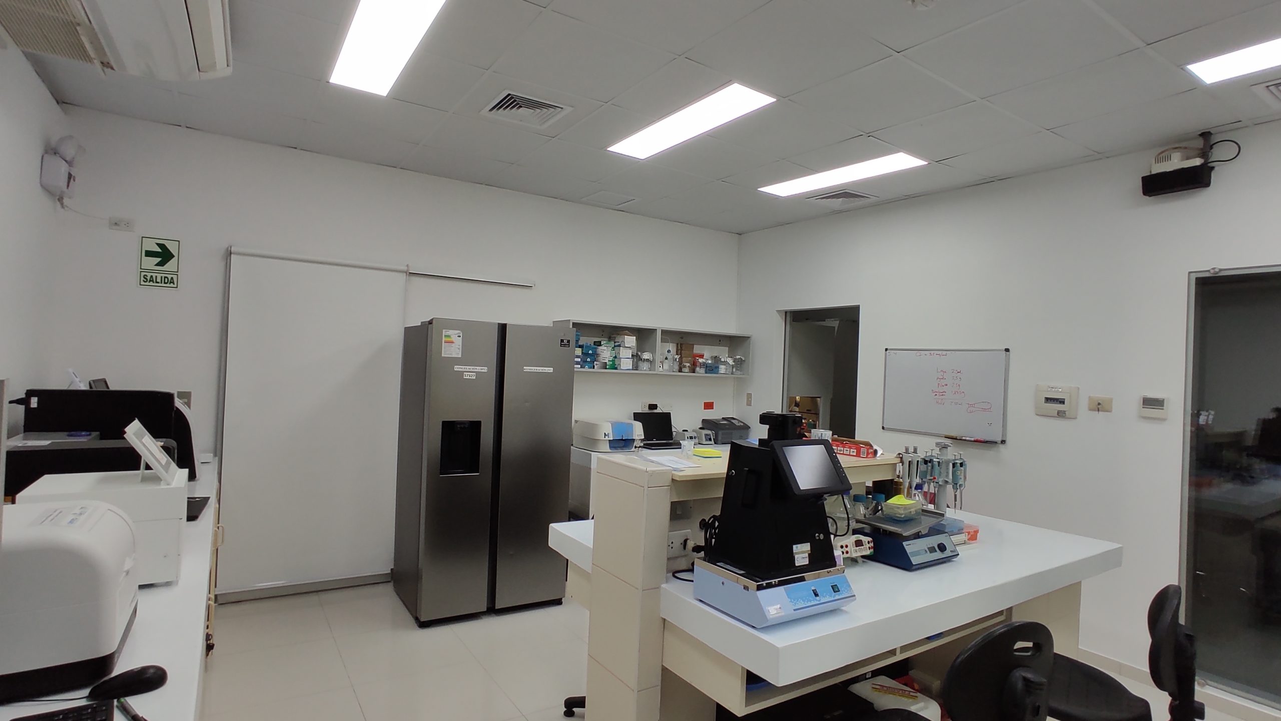
Equipamiento

MAGPIX – Analizador Multiplex de Biomarcadores Genomicos
Purificador de Agua
Liofilizador
Balanza Analitica
Centrifuga Digital
Camara de Electroforesis con fuente de poder
Microscopio Invertido
Cabina de Bioseguridad Clase II
Contador Celular Automatizado Portatil
Separador Celular Magnetico
Lector de Microplacas
Cabina de Flujo Laminar
Lector Multimodal de Microplacas
QNANO – TRPS – Analizador de Nanoparticulas con Deteccion de Pulso Resistivo Sintonizable
Medidor de LUX PC
Termociclador PCR Tiempo Real
Incubadora CO2
Centrifuga Refrigerada
Secuenciador de ADN
Sistema de Documentacion de Geles
Microscopio de Epifluorescencia con Camara

Capacidades

  • Búsqueda de compuestos bioactivo derivados de plantas medicinales o macroalgas mediante el uso de cultivo de células cancerosas
  • Aislamiento de células madre de diferentes especies de animales con fines preclínicos
  • Aislamiento de células madre del cáncer de líneas celulares para el análisis de exosomas

Colaboración (Instituciones)

Maria Lucia Zaidán Dagli, DVM, PhD. Escuela de Medicina Veterinaria y Ciencias Animales, Universidad de São Paulo, Brasil.
Timothy Thomson Okatsu, MC, PhD. Instituto de Biología Molecular de Barcelona. CSIC, España.
Carlos Castañeda Altamirano, MC, MSc. Oncólogo. Instituto Nacional de Enfermedades Neoplásicas. Lima, Perú.
José González Cabeza, Bgo, PhD. Grupo de Investigación de Microbiología Molecular y Biotecnología de la Facultad de la Ciencias de la Salud de la UPAO. Trujillo, Perú.
Cristian Torres Mendoza, DVM, PhD. Laboratorio de Biomedicina y Medicina Regenerativa. Departamento de Ciencias Clínicas. Facultad de Ciencias Veterinarias y Pecuarias. Universidad de Chile. Santiago, Chile.
Yanina Panzera (PhD), Sección Genética Evolutiva, Universidad de la República, Montevideo, Uruguay.
Obert Marin Sanchez. Mg. Universidad Nacional Mayor de San Marcos. Peru.
Alexis German Murillo Carrasco. Dr. Universidad de Sao Paulo. Brasil.
Alexei Vicent Santiani Acosta, Dr. Universidad Nacional Mayor de San Marcos.
Tatiana Vidaurre, Dra. Instituto Nacional de Enfermedades Neoplásicas. Lima, Perú.
Alfredo Aguilar, Dr. Oncosalud. Lima, Perú.
Libertad Alzamora Gonzales, Dra. Grupo de investigación: Inmunoestimulantes de origen natural. Facultad de Ciencias Biológicas. Universidad Nacional Mayor de San Marcos. Lima, Perú.

Links de interés

Laboratorio de Investigación en Cultivo Celular e Inmunología Molecular
Procedimiento para el cultivo e identificación de células madre obtenidas de lipoaspirado humano con fines de investigación

Las células madre humanas nacen con la creación de la vida misma y algunas de estas permanecen durante toda la vida. Por consiguiente, se pueden hallar en tejidos adultos y utilizarlas para investigaciones a nivel básico y aplicado. Actualmente, en nuestro país existe un creciente interés en el estudio y aplicación de células madre; sin embargo, existe poco conocimiento acerca del procedimiento para su identificación. Es por ello que este artículo tiene como objetivo dar a conocer, desde un punto de vista práctico, un procedimiento para el cultivo e identificación de células madre/estromales obtenidas de lipoaspirado humano (Adipose Stem Cells) con fines de investigación, el cual incluye la caracterización a nivel de inmunofenotipo, el potencial de diferenciación celular, la expresión génica y el control de calidad del cultivo celular, que sirva de apoyo para los profesionales de la comunidad científica peruana que deseen desarrollar esta línea de investigación.

Laboratorio de Investigación en Cultivo Celular e Inmunología Molecular
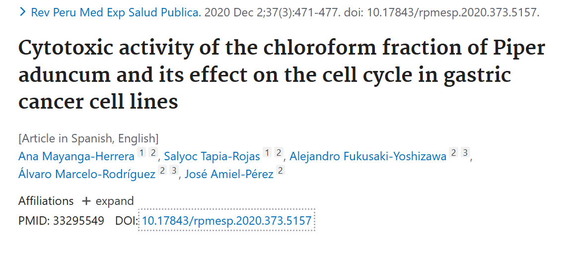
Cytotoxic activity of the chloroform fraction of Piper aduncum and its effect on the cell cycle in gastric cancer cell lines

Objectives: To evaluate the cytotoxic activity of the chloroform fraction of the Piper aduncum methanolic extract (PAMoCl) and its effect on the cell cycle in two gastric cancer cell lines: AGS and KATO III.

Materials and methods: The cytotoxic effect of PAMoCl was evaluated in cell lines AGS and KATO III. The following PAMoCl concentrations were tested, 1.25, 2.5, 5, 10, 20, 40, 80 and 160 μg/mL. Resazurine was used to evaluate cell viability. In the cell cycle assay, the cells were treated with 19.62 μg/mL and 39.23 μg/mL of PAMoCl for AGS as well as 87.49 μg/mL and 160 μg/mL for KATO III. After 24 hours both cell lines were analyzed by flow cytometry.

Results: PAMoCl showed cytotoxic activity, inhibiting cell growth by 50%. It presented a (IC50) of 39.23 μg/mL and 87.49 μg/mL at 24 hours and a (IC50) of 49.47 μg/mL and 64.68 μg/mL at 48 hours against AGS and KATO III cell lines, respectively. In addition, it was observed that PAMoCl has an effect on the cell cycle, it causes an accumulation of cells in the G2/M phase.

Conclusions: PAMoCl contains secondary metabolites with cytotoxic activity that have an effect on the G2/M phase of the cell cycle, in two gastric cancer cell lines, both primary and metastatic. The results of this study will allow us to deepen the search for more effective active ingredients found in PAMoCl for eliminating gastric cancer cells, but with less toxicity for healthy cells.

Laboratorio de Investigación en Cultivo Celular e Inmunología Molecular
Cytotoxicity of Fractions of Dracontium spruceanum Chloroform Extract in MDA-MB-231 and MCF-7 Breast Cancer Cell Lines

Background:

Breast cancer is a major health issue responsible for numerous deaths worldwide. Medicinal plants are rich in compounds with potential anticancer properties.

Objectives:

This study aimed to assess the cytotoxicity of fractions from the Dracontium spruceanum chloroform extract on MDA-MB-231 and MCF-7 breast cancer cell lines.

Methods:

Eleven fractions were screened for cytotoxic effects on cell viability using the MTT assay. Subsequently, the two most cytotoxic fractions were selected, and the half-maximal inhibitory concentration (IC50) was calculated using two-fold dilution concentrations ranging from 100 to 1.56 µg/mL.

Results:

Fractions F43 and F50 showed the highest cytotoxicity at 100 µg/mL. The IC50 of F43 was 270.8 ± 18.07 µg/mL and 133.0 ± 17.99 µg/mL for MDA-MB-231, and 252.2 ± 21.94 µg/mL and 144.3 ± 23.14 µg/mL for MCF-7 cells at 24 and 48 hours, respectively. For F50, IC50 values were 107.2 ± 2.97 µg/mL and 126.1 ± 14.77 µg/mL for MDA-MB-231, and 236.4 ± 58.7 µg/mL and 104.5 ± 10.54 µg/mL for MCF-7 at corresponding times.

Conclusions:

 Dracontium spruceanum fractions demonstrated moderate cytotoxicity on MDA-MB-231 and MCF-7 breast cancer cell lines, indicating their anticancer potential. Further research is required to identify the metabolites responsible for this activity and to elucidate the molecular mechanisms involved.

Laboratorio de Investigación en Cultivo Celular e Inmunología Molecular
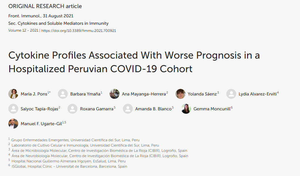
Cytokine Profiles Associated With Worse Prognosis in a Hospitalized Peruvian COVID-19 Cohort

Cytokines, chemokines and growth factors present different expression profiles related to the prognosis of COVID-19. We analyzed clinical parameters and assessed the expression of these biomarkers in patients with different disease severity in a hospitalized Peruvian cohort to determine those associated with worse prognosis. We measured anti-spike IgG antibodies by ELISA and 30 cytokines by quantitative suspension array technology in 123 sera samples. We analyzed differences between patients with moderate, severe and fatal COVID-19 by logistic regression at baseline and in longitudinal samples. Significant differences were found among the clinical parameters: hemoglobin, neutrophils, lymphocytes and C-reactive protein (CRP), creatinine and D-dimer levels. Higher anti-spike IgG antibody concentrations were associated to fatal patient outcomes. At hospitalization, IL-10, IL-6, MIP-1α, GM-CSF, MCP-1, IL-15, IL-5, IL1RA, TNFα and IL-8 levels were already increased in fatal patients´ group. Meanwhile, multivariable analysis revealed that increased GM-CSF, MCP-1, IL-15, and IL-8 values were associated with fatal outcomes. Moreover, longitudinal analysis identified IL-6 and MCP-1 as the main risk factors related to mortality in hospitalized COVID-19 patients. In this Peruvian cohort we identified and validated biomarkers related to COVID-19 outcomes. Further studies are needed to identify novel criteria for stratification of SARS-CoV-2 infected patients at hospital entry.

Galería multimedia

Conoce la actividad del laboratorio aquí.